Introduction
- Entamoeba hartmanni is a non-pathogenic amoeba with world wide distribution. Its life cycle is similar to that of E. histolytica but it does not have an invasive stage and does not ingest red blood cells.
Morphology of Trophozoites
- Morphology of the trophozoites is similar to those of E. histolytica / dispar but they do not contain ingested red blood cells and the motility is less rapid.

Entamoeba hartmanni trophozoite wet mount preparation, notice the extending pseudopods extending pseudopods in multiple directions and the karyosome here is eccentrically located.
Morphology of Cysts
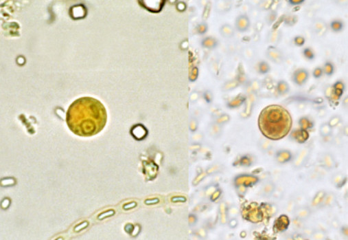
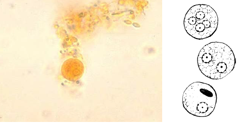
- Cysts of E. hartmanni 7-9μm in diameter and contain one to four nuclei. Chromatoid bodies are usually present in young cysts as elongated bars with bluntly rounded ends. Glycogen is usually diffuse, but in young cysts it is often present as a concentrated mass, staining reddish brown with iodine.

Entamoeba hartmanni cysts Iodine stain – Left, a mononucleate cyst with a visible chromatoid body. Right, a mononucleate cyst with a well-stained glycogen vacuole.
References:
- CDC
- http://www.atlas-protozoa.com


