
- Before we start with the abnormal morphologies, let’s talk about normal morphology of Red Blood Cells.
- Normal mature RBC are biconcave, round discs that are about 6 – 8 in diameter, which is only slightly smaller than the normal small mature lymphocytes ( about 6 – 10 in diameter).
- The term used to indicate red blood cells of normal size and shape is normocytic. The term used to indicate a normal color or central pallor (i.e., normal hemoglobin content) is normochromic

Normocytic and Normochromic RBC
Hypochromic: Erythrocytes that demonstrate a central pale area that becomes larger and paler as the hemoglobin content diminishes.
- Iron Deficiency
- Sideroblastic Anaemia
- Thalassaemia

Hypochromic RBC
Anisochromic: indicates the presence of both normochromic and Hypochromic

Anisochromic (Normochromic plus Hypochromic)
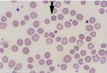
Polychromasia: Changeable terms used to indicate the increased presence of non-nucleated immature erythrocytes (Polychromatophilic erythrocytes) that contain residual RNA which gives a blue-gray tint to the red cells. These cells, which remain after ejection of the nucleus from the orthochromatic erythroblast are slightly larger than mature erythrocytes. After exposure to a supravital stain, the cytoplasm organelles of these cells clump into an easily recognized blue-staining reticulum and the cells is called a reticulocytes.
Polychromasia
Anisocytosis: is a “generic” term used to indicate variation in shape of erythrocytes (e.g. oval, pear-shaped, teardrop-shaped, saddle-shaped, helmet-shaped, sickle-shaped, and irregularly shaped).

Anisocytosis
Microcytosis: abnormally small erythrocytes (i.e., less than 6 in diameter). compare with the size for small lymphocyte.
- Found with Hypochromic

Microcytosis
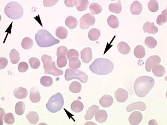
Macrocytosis: abnormally large erythrocytes (i.e., less than 8 in diameter).
- Megaloblastic Anaemia
- High reticulocytes count
- Liver disease
- Myelodysplastic syndrome

Macrocytosis
Target Cells (Codocytes): erythrocytes that are thinner than normal which show a peripheral rim of hemoglobin with a dark central hemoglobin-containing area. A pale unstained ring containing less hemoglobin separates the central and peripheral zones and gives the cell a target appearance.
- Liver Disease
- Hemoglobinopathies
- Thalassaemia
- Sideroblastic anemia

Target Cells
Spherocytes: are nearly spherical erythrocytes which are nearly spherical erythrocytes which usually have a diameter smaller than normal. They lack the central pale area due to their spherical shape.
- Hemolytic anemia
- Post transfusion
- Hereditary sphercocytosis
Spherocytes
Tear-Drop Cells:
- Severe Anaemia
- Myloproliferative disorders

Teardrop cells (dacrocytes)
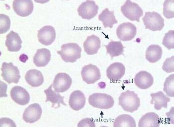
Elliptocytes and Ovalocytes: are interchangeable terms used to indicate ovalshaped erythrocytes.
- Hereditary elliptocytosis
- Iron-deficiency anaemia
- Thalassaemia

Elliptocytes
Stomatocytes:
- Acute alcoholism
- Malignancies

Stomatocytes
Helmet Cells:
- G6PD deficiency
- Pulmonary emboli

Helmet Cells

Helmet Cells (Keratocytes)
Schistocytes: are fragmented red cell segments that are the result of some hemolytic process. These segments can be a variety of shapes but helmet cells and triangularly-shaped cells are particularly characteristic.
- Disseminated intra-vascular coagulopathy (DIC)
- Thrombotic Thrombocytopenia purpura (TTP)
- Hemolytic uremic syndrome (HUS)

Schistocytes
Sickle cells (drepanocyes): are interchangeable terms used to indicate sickle-like forms of erythrocytes (crescent-shaped, irregular spines, filaments, holly-leaf appearance) noted when RBC containing HbS are subjected to reduction in oxygen tension or pH.
- Sickle cell anaemia
- Sickle thalassaemia

Sickle Cells
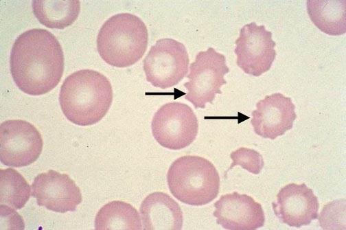
Acanthocytes (Spur Cell):
- Congenital abetalipoproteinemia
- Vitamin E deficiency
- Alcohol intoxication
- Post-splenectomy
Acanthocytes
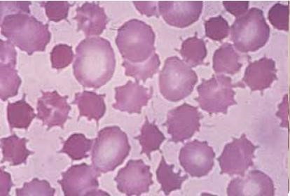
Burr Cells:
- Liver disease
- Renal disease
- Severe burns
- Bleeding gastric ulcers
- Maybe artifact
Echinocyte (Burr Cell)
Acanthocyte vs Echinocyte
Howell-Jelly: are intracellular particles which are smooth, round remnants of nuclear chromatin (DNA. Usually, only one per cell is seen but, occasionally, there may be more than one
- Megaloblastic anaemia
- Mylodysplastic

Howell-Jolly Bodies
Heinz Bodies

Heinz Bodies
Cabot Rings

Cabot Rings
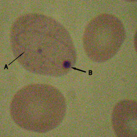
A – Cabot ring
B – Howell-Jolly body
Basophilic Stippling: is the term used to indicate the presence of irregular basophilic granules in the cytoplasm of erythrocytes. The granules are composed of unstable RNA and may be fine of coarse.
- Lead Poisoning
- Thalassaemia
- Significant anaemia
- Dyserythropoiesis

Basophilic Stippling
Hemoglobin C crystals: are hexagonal crystals that may be found in individuals with HbC syndromes. The crystals may intracellular or extra-cellular.

Hemoglobin C crystals
Pappenheimer Bodies: are intracellular inorganic iron-containing granules that may be ob-served on Wright’s stained peripheral blood smears.

Pappenheimer Bodies
Rouleaux formation: describes an aggregation of erythrocytes that are aligned one upon the other, resembling stacks of coins
- High level of circulating acute-phase proteins.
- with High ESR rate.
- autoimmune conditions
- myeloma
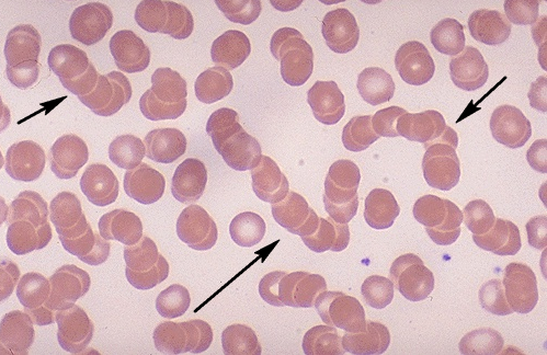
Rouleaux formation
Agglutination of red cells: is caused by agglutinins and resembles Rouleaux but is more irregular with round clumps rather than linear Rouleaux

Agglutination of red cells
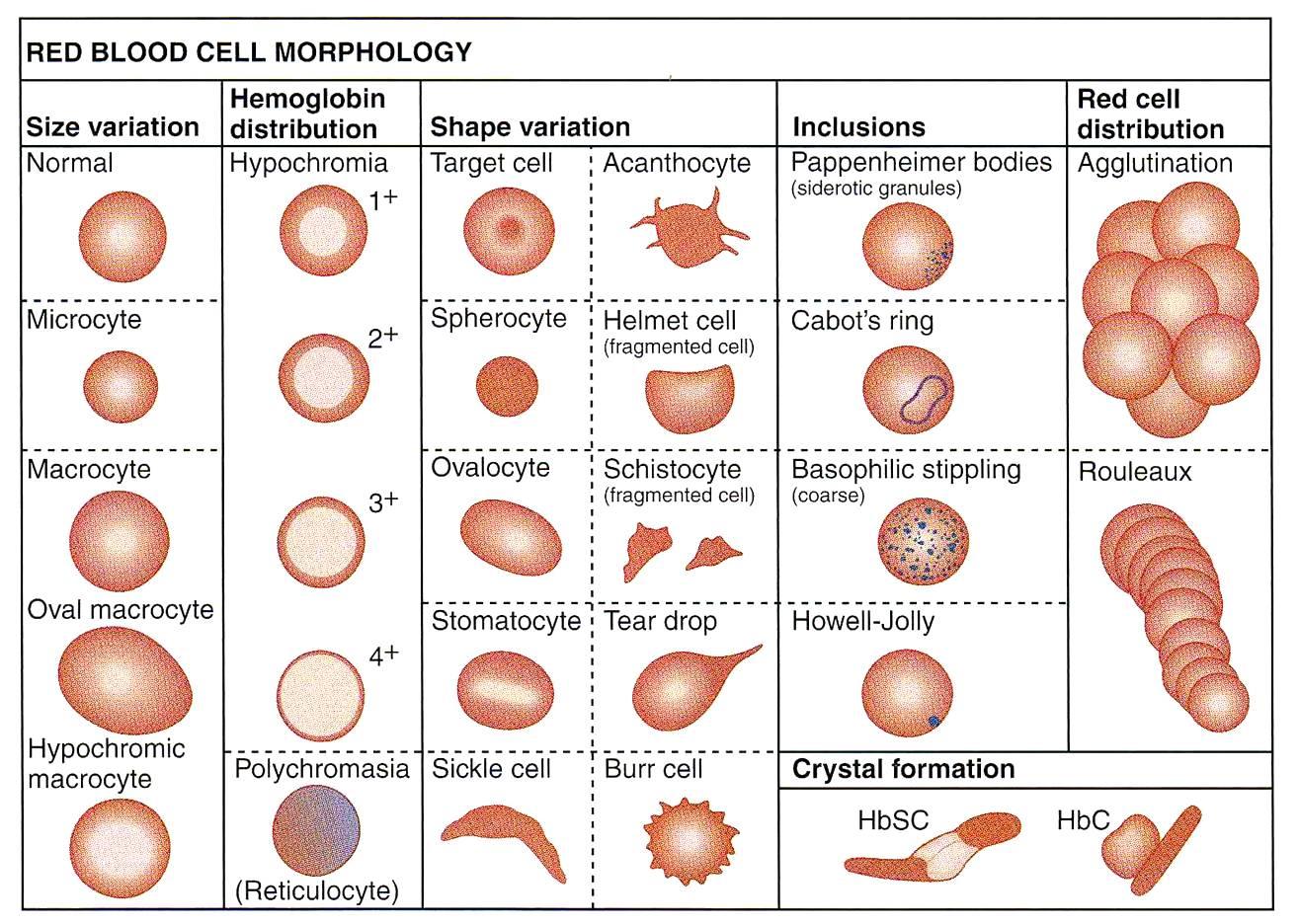
Summary of red blood cellsmorphology