Introduction
Trichuris trichiura, more commonly known as the Whip Worm, due to the whip-like form of the body. These nematodes are most commonly seen in tropical climates and in areas where sanitation is poor. They seem to occur in areas particularly where Ascaris and Hookworms are found due to the eggs requiring the same conditions to allow for embryonation. Both species can be found in humans together. There are several species within this genus each infecting specific hosts, but only T. trichiura infects man. Causing human trichuriasis. It is a parasite that infects many more people than is generally appreciated, up to 800 million people throughout the tropics and temperate regions.
Life cycle
eggs require a warm, moist environment with plenty of oxygen to ensure embryonation, but once they have embryonated they are extremely resistant to environmental conditions. Adult worms are found in the cecum and upper part of the colon of man. In heavy infection they can be found in the colon and the terminal ileum. They attach to the mucosa by the anterior end or by embedding the anterior portion of the body in the superficial tissues, obtaining nutrition from the host tissues.
Once fertilized the female worms lay several thousands of eggs, which are unsegmented at the oviposition and are passed out in the feces. Once they have been passed out they require an embryonation period in the soil which may last from two weeks to several months, after which they become infective. When embryonated eggs are swallowed by human hosts larvae are released into the upper duodenum. They then attach themselves to the villi lower down the small intestine or invade the intestinal walls. After a few days the juveniles migrate slowly down towards the cecum attaching themselves to the mucosa, reaching their final attachment site simultaneously.
The larvae reach maturity within three weeks to a month after infection, during which they undergo four molts. There is no lung migration and the time from ingestion of infective eggs to the development of adult worms is about three months. Infection is achieved by swallowing soil that contains embryonated eggs. Therefore, children are most commonly seen to possess the infections, as they are more likely to swallow soil.
Morphology
The adult worms of T. trichuria are characterized by the enormously elongated capillary-like esophagus (anterior end); with the anus situated in the extreme tip.
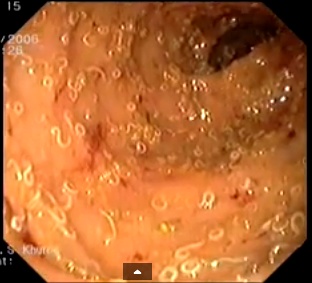
The thin anterior portion of the worm is found embedded in the mucosa. There are no lips and the vulva is at the junction of the thread-like and thickened regions of the body. The posterior end is much thicker and lies free in the lumen of the large intestine.
The female measures 35-50mm long and the male 30-45mm long.
The ova are characteristically barrel shaped, bile stained with bipolar plugs. They measure 50-54μm by 20-23μm.
Clinical Disease
Most infections due to this nematode are light to moderate with minimal or no symptoms. However, a heavy worm burden may result in mechanical damage to the intestinal mucosa due to the adult worm being threaded into the epithelium of the cecum. Abdominal cramps, tenesmus, dysentery and prolapsed rectum may occur in these cases.
If a prolapsed rectum is observed, many worms may be seen adhering to the mucosa of the rectum. Symptomatic infections are usually only seen in children. The majority of infections are chronic and mild, with nonspecific symptoms like diarrhea, anemia, growth retardation, eosinophilia.
Laboratory Diagnosis
The adult worms of T. trichiura are rarely seen in the feces. The microscopic examination of stool deposits after an iodine stained, formol-ether concentration method concentration reveals the characteristic barrel shaped ova. In symptomatic infections numerous numbers of eggs can be seen due to the prolific nature of the female worms, even in light infections many eggs can be seen in the smear.


